Confocal Microscopy and Flow Cytometry Laboratory
Function of the infrastructure
The Confocal Microscopy and Flow Cytometry Laboratory occupies is a vital apparatus of the centre Armand-Frappier Santé et Biotechnologie. Used by multiple teams for a large variety of projects, this research infrastructure offers the necessary tools to complete projects using flow cytometry protocols, as well as giving access to a confocal microscope and the expertise of the platform managers for proper use of the provided tools and equipment.
Flow cytometry is a technique that aims to quantify and measure cells from a sample. In its simplest form, a cytometry protocol could be completed using an optical microscope, from which the user could count manually previously coloured cells. While flow cytometry is based on this simple principle, it also rests on the back of numerous technological advances that allow the automatisation of the precess as well as notably increased accuracy.
The flux cytometre measures the interaction between one or multiple light sources (usually lasers) and a liquid flow that canalizes and conveys the cells in a single file. When cells pass through the laser, the emitted light will be diffracted according to the size and the morphology of the affected cell, producing various signals that can be amplified and measured by the optical system (composed of dichroic mirrors and light detectors) and converted in electrical signals which can then be interpreted by a linked computer. Some protocols will also need the addition of fluorochrome antibodies specific to cellular markers of interest, which will then be excited when exposed to the laser and emit photons which will also be captured by the optical system.
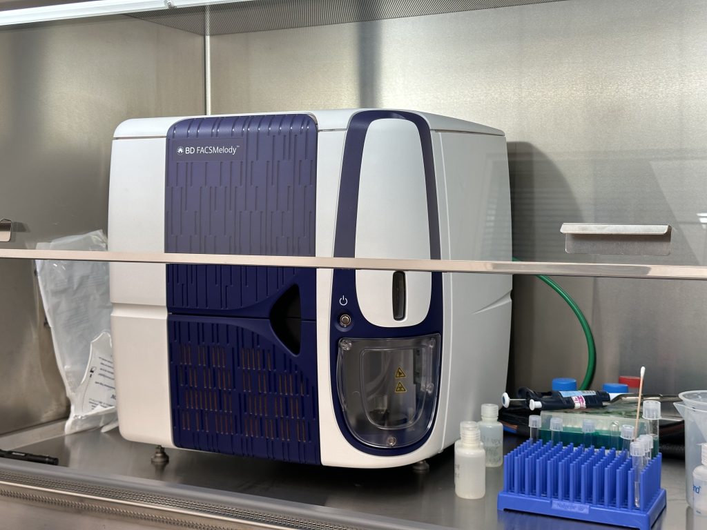
Cytomètre en flux FACSMelody de BD qui possède une fonction de trieur de cellules, sous une hotte à flux laminaire pour permettre les expériences de culture cellulaire stériles
Data gathered by such protocols includes light scattered at small angles (forward scatter, which is correlated to the size of the cell), light scattered at 90 degrees (side scatter, which varies according to granularity and interior components) and emitted fluorescence (autofluorescence produced by cells as well as fluorescence coming from excited fluorochromes). These results are displayed as multi or mono-parametric histograms which describe the intensity of the captured light for multiple parameters, which can then be used to identify the various possible types of cells contained within a sample, contaminants and diverse other factors.
While a majority of flux cytometers currently available on the market allow only one reading of a sample, being unable to be reused, some models offer a function of cell sorting which allows the isolation of cells according to the captured signals, thus grouping together various cell populations in separate containers. The flow, after passing through the optical system, is broken by a vibrating nozzle at a specific wavelength, allowing the formation of droplets that contain single cells. These droplets, at the moment they break off the flow, obtain an electromagnetic charge according to the identity the computer assigned it, through the activity of an electromagnetic charging ring positioned at the tip of the nozzle. This charge allows the triage by a magnetic current which will deviate the trajectory of the particule according to its charge, guiding the droplet towards the proper container. These cells can then be reused in subsequent experiments, or simply be recultivated with the confidence of having a pure uncontaminated sample of a single cell type.
A confocal microscope differs from an optical microscope by its production of a virtual three-dimensional representation of the studied object, rather than an enlargement of the object through the use of lens that can be observed with the naked eye.
The confocal microscope was created as a means to nullify limitations of traditional optical microscopes that get worse as magnification increase, such as a difficulty to obtain clear pictures of thick or high relief subjects which leave the focal point of the microscope. Additionally, traditional optical microscopes flood the whole subject with light, which can cause problems during fluorescence experiments, where signals coming from outside of the focal plane are blurry and can reduce simultaneously the clarity of the picture and of signals that are coming from the focal plane.
These problems are avoided by two important changes: illumination comes from a laser that will brighten the subject in a point-by-point manner, and the reflected light must pass through an aperture before being detected, which prevents the passage of light or fluorescence from outside of the focal plane of the lens. Upon illumination, the signal will thus pass through the aperture and then be amplified by photomultipliers and stored as electric currents varying in intensity according to the strength of the captured signal. A new point will then be illuminated, until the entire pre-determined surface area has been covered by the apparatus. Afterwards, a picture can be produced by a computer using the data obtained during the scan, creating a visual representation of the captured signals at varying positions on the Z-axis.
In order to create three-dimensional models, movement on the Z axis is needed in order to put a new layer of the subject in the focal plane of the lenses. Upon layering the pictures together, a 3D model of the subject is produced, where each picture illustrates a thin layer of the studied object. Pictures can also be captured after successive time lapses, allowing the production of videos that show changes over time in living models.
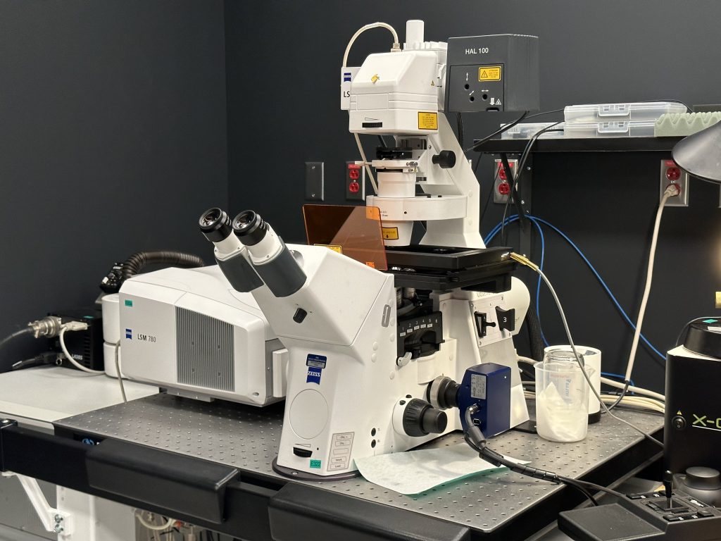
Confocal microscope LSM 780 by Zeiss
Example of a time lapse visualisation using confocal microscopy. Two communal parasittrophic vacuoles fused in order to produce a bigger one. Model using bone marrow-derived macrophages, obtained from transgenic Life-Act mice that are engineered to allow visualisation of polymerized actin. These cells are aditionally infected by Leishmania Amazonensis GFP.
Video produced by Christine Matte
The laboratory and the Infectiopole
This research infrastructure favorises the progress of a variety of projects that use cellular models. The various uses of flow cytometry allow staff to identify the components of a cell populations, as well a sort them according to the needs and characteristics valued by the research protocol. Additionally, the confocal microscope can be used in order to visualize cells in a variety of contexts, without being impaired by the limitations of optical microscopy.
Instruments
- A BD FACSMelody highly automated cell sorter that is easy to learn and allows the isolation of up to 4 different cell types in a single sample.
- A confocal microscope LSM 780 by Zeiss.
Contact information
Jessy Tremblay Terms of use and pricing
- jessy.tremblay@inrs.ca
- +1 (450) 687-5010, poste 4314